

Welcome to Oyster Anatomy 101: The In’s and Out’s, a comprehensive guide designed to explore the intricate world of oyster anatomy. This article delves into the fascinating structural and functional aspects of oysters, breaking down their anatomy into three essential sections for a clear and detailed understanding. First, we will examine the basic anterior and posterior anatomy, focusing on the shell’s external features and their roles. Next, we’ll explore the digestive system, highlighting how oysters process and absorb nutrients from their environment. Finally, we’ll uncover the internal anatomy, including the critical organs responsible for reproduction, respiration, and waste management. Whether you’re a marine biology enthusiast or a curious learner, this guide will provide valuable insights into the remarkable adaptations that allow oysters to thrive in their aquatic habitats.
Basic Oyster Anatomy: Anterior & Posterior
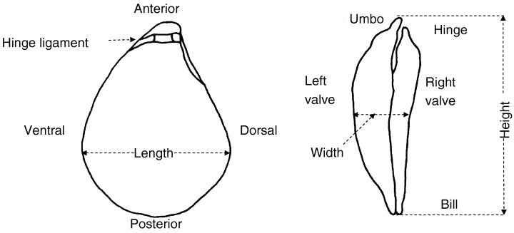
Before you dive into internal organs and digestive pathways, it helps to understand how the oyster shell itself is laid out. These key landmarks—hinge, umbo, valves, and shell edges— define the oyster’s orientation and how it opens, closes, and feeds.
Reference: Martin, D. (2006). Oyster shucking technologies: Past and present. International Journal of Food Science & Technology, 41.
External Shell Landmarks
Location: Positioned along the dorsal side, connecting the two shell valves near the umbo.
Function: A flexible, elastic band that works like a spring. When the adductor muscle relaxes, the hinge ligament helps the shell open; when the muscle contracts, the valves close tightly to protect the oyster.
Location: The rounded, raised area near the hinge on the dorsal side of the shell.
Function: The oldest part of the oyster shell and the initial point of shell growth. It provides structural integrity and helps identify the dorsal side during shucking and scientific orientation.
Location: The larger, deeper valve that normally sits on the bottom when the oyster is in its natural resting position.
Function: Provides a stable base and often attaches to rock, shell, or farm gear. The left valve protects the oyster’s soft body from predators and environmental stress.
Location: The flatter, thinner valve that closes over the left valve.
Function: Acts like a lid, closing firmly over the left valve to fully enclose and safeguard the oyster’s body against impact, drying, and predators.
Location: The thin, elongated edge of both valves opposite the hinge, forming the anterior end of the oyster.
Function: Allows the oyster to open slightly for filter feeding and respiration. The bill helps direct water and suspended food particles into the shell.
Location: The lower edge of the oyster, opposite the dorsal side where the hinge and umbo sit.
Function: The main opening edge where the valves separate the widest during feeding and respiration, allowing water flow through the gills.
Location: The upper side of the oyster where the hinge ligament and umbo are located.
Function: Provides structural support and houses the hinge apparatus responsible for opening and closing the shell.
Anterior side: The “front” of the oyster, generally where the bill is located. Internally, this region contains the mouth and labial palps.
Posterior side: The “back” of the oyster, closer to the hinge and umbo. Internally, this is typically where the anus and excurrent siphon are found.
Shell Function Overview
Protection: The left and right valves, reinforced around the umbo and hinge, shield the oyster’s soft body from predators, wave action, and environmental stress.
Feeding: The bill and ventral side allow the shell to gap just enough for water to flow across the gills, where food particles are captured and sorted.
Movement of the Shell: The hinge ligament and adductor muscle work together as a spring-and-clamp system, controlling how and when the shell opens or closes.
Support & Orientation: The dorsal side and umbo give structural integrity and a clear “up” direction, helping shuckers, farmers, and researchers consistently orient the oyster.
Digestive Oyster Anatomy
An oyster’s digestive system is compact but efficient. From the esophagus to the rectum, each organ plays a specific role in sorting, breaking down, absorbing, recycling, and finally expelling food and waste.

Reference: Prasetiya, F. (2015). Greening phenomenon in bivalve by marennine produced from Haslea ostrearia and its consequences on bivalve’s integrated response.
Overview: How an Oyster Processes Food
Oysters are filter feeders, pulling water across their gills, trapping tiny particles, and sending selected food into a tight loop of organs packed into the visceral mass. Once particles are accepted as “food,” they move from the mouth and esophagus into the stomach, where enzymes from the digestive gland and crystalline style begin to break everything down. From there, the gut loops through the body, stripping out usable nutrients before compressing what is left into compact waste pellets.
Key Digestive Organs and Their Roles
While the diagram shows a tangle of tubes and sacs, each part has a clear job. The organs below work together to move food, apply enzymes, absorb nutrients, and finally package and expel waste without wasting much energy or space inside the shell.
Function: A short tube that connects the mouth to the stomach, transporting food particles after they are sorted by the labial palps.
Function: A sac-like chamber where food first meets digestive enzymes from the digestive gland and crystalline style, beginning chemical breakdown.
Function: Surrounds the stomach and produces digestive enzymes. It also absorbs, processes, and stores nutrients from digested food.
Function: The style sac houses the crystalline style, a rod of digestive enzymes that rotates and grinds food. The midgut is the main site of enzymatic digestion and nutrient absorption.
Function: Carries digested material downward from the midgut, continuing the process of digestion and nutrient absorption.
Function: Carries partially digested material back toward the stomach region, allowing further breakdown and nutrient extraction before waste formation.
Function: Final section of the digestive tract where undigested material is compacted and prepared for excretion as fecal pellets.
Function: Terminal opening of the digestive system, located near the excurrent siphon, where compacted waste is expelled from the oyster’s body.
Digestive Process Overview
From the first capture of a food particle to the final expulsion of waste, oyster digestion follows a clear sequence that maximizes nutrient extraction from filtered water.
Want more chef insight? The Raw Bar Bible has it all.
The Raw Bar Bible
A deeper look into oysters, shucking technique, brine, knife choice, and real raw bar workflow — written from a chef’s point of view. Clear, practical, and built from actual service experience.
Overall Internal Oyster Anatomy
Inside the shell, oysters pack a surprising amount of anatomy into a small space. These core organs manage filtration, digestion, reproduction, circulation, and water flow within the mantle cavity.

Location: At the anterior end of the visceral mass, just behind the labial palps.
Function: Receives sorted food from the palps and channels it through a short esophagus into the stomach.
Location: Central visceral mass, surrounded by the digestive gland.
Function: Mixes incoming food with digestive enzymes from the gland and crystalline style to begin chemical breakdown.
Location: Surrounds the stomach in the central visceral region.
Function: Produces digestive enzymes and absorbs, stores, and processes nutrients from digested food.
Location: Extends from the stomach into the midgut.
Function: Holds the crystalline style, a rotating rod that grinds food and releases digestive enzymes into the gut.
Location: Loops through the visceral mass and often over the adductor muscle.
Function: Continues enzymatic digestion and absorbs remaining nutrients as material moves toward the rectum.
Location: Near the posterior region, close to the excurrent siphon.
Function: Releases compacted waste into the excurrent water stream for removal from the shell.
Location: On either side of the mouth, adjacent to the gills.
Function: Sort and select suitable food particles captured by the gills and direct them to the mouth.
Location: Near the heart, beneath the pericardium.
Function: Filters waste from hemolymph and helps regulate internal fluid and salt balance.
Location: Dorsal side of the body, within the pericardial cavity.
Function: Pumps hemolymph through the oyster’s body, supplying tissues with oxygen and nutrients.
Location: Membranous sac surrounding the heart on the dorsal side.
Function: Protects the heart and helps maintain proper hemolymph pressure around it.
Location: Surrounds much of the digestive gland and intestine.
Function: Produces eggs or sperm; expands and ripens during the spawning season.
Location: Small branching channels extending from the gonads and primary ducts.
Function: Carry reproductive cells toward the mantle cavity and assist in moving some waste products outward.
Location: Soft tissue lining the interior of both shell valves.
Function: Secretes shell material, participates in gas exchange, and helps expel certain wastes into the mantle cavity.
Location: Space between the mantle and visceral mass.
Function: Houses the gills and provides the main chamber where water flows for feeding, respiration, and waste removal.
Location: On both sides of the body, projecting into the mantle cavity.
Function: Extract oxygen from water, capture food particles, and help regulate water movement through the shell.
Location: Ventral/posterior opening along the mantle edge.
Function: Draws fresh, particle-rich water into the mantle cavity for feeding and gas exchange.
Location: Near the posterior/dorsal region, close to the anus.
Function: Expels used water, waste, and gametes out of the shell.
Location: Large central muscle attaching both valves internally.
Function: Closes the shell tightly against predators and desiccation; relaxes to allow partial opening for feeding.
You Might Also Want to Read
Frequently Asked Questions About Oyster Anatomy
Functional Overview of Key Oyster Organs
The organs below work together to keep an oyster feeding, breathing, reproducing, and regulating its internal environment.
-
KidneyFiltration & Balance
- Filters metabolic waste from the hemolymph and helps regulate internal salinity (osmoregulation), keeping the oyster’s internal fluids in balance with surrounding water.
-
Crystalline Style SacEnzymatic Rod
- Houses the crystalline style, a rotating rod of digestive enzymes that grinds and dissolves food as it passes through the stomach and midgut, boosting chemical digestion.
-
Secondary DuctsTransport Network
- Fine branching channels that move reproductive cells and assist in routing certain waste products from deeper tissues toward the mantle cavity and surrounding water.
-
Labial PalpsFood Sorting
- Sit beside the mouth and act as the oyster’s sorting station—accepting suitable food particles brought in by the gills and rejecting excess or unsuitable material as pseudofeces.
-
GonadReproduction
- Produces eggs or sperm and fills much of the visceral mass during the spawning season. Its texture and color shift as gametes mature and are released into the water.
-
PericardiumHeart Cavity
- A thin, protective sac that surrounds the heart on the dorsal side of the body, helping maintain the right pressure and fluid environment for effective hemolymph circulation.
-
MantleShell & Interface
- Lines the inner surface of the shell, secreting new shell layers, participating in gas exchange, and helping route wastes and gametes into the mantle cavity and out through the siphons.
-
GillsBreathing & Feeding
- Large, folded sheets of tissue that drive water flow, extract oxygen, and capture tiny food particles. Cilia on the gills move mucus-bound particles toward the labial palps for sorting.
General Overview
General Overview
The anatomy of an oyster is a highly efficient system built to support life as a filter feeder. From the protective shell to the intricate internal organs, each structure contributes to feeding, respiration, reproduction, and survival in dynamic marine environments.
External Structure: The Oyster’s Protective Framework
The outer anatomy includes the left and right valves, hinge ligament, and umbo. These structures allow the oyster to anchor securely, regulate shell opening for feeding and respiration, and channel water flow across the gills. The dorsal and ventral regions, along with the bill, help maintain orientation and optimize filtration efficiency.
Internal Systems: How the Oyster Lives, Breathes, and Feeds
Beneath the shell is a coordinated array of organs responsible for digestion, respiration, circulation, and reproduction. Together they form a compact but highly effective biological system.
Digestive System
The esophagus, stomach, digestive gland, intestines, rectum, and crystalline style sac work in sequence to break down and absorb nutrients from filtered particles. The labial palps ensure only suitable food reaches the mouth, maximizing energy efficiency.
Reproductive System
The gonads and secondary ducts support gamete production and release. Because oysters spawn externally, this system plays a vital role in population survival and genetic diversity.
Waste Removal & Circulation
The kidney filters metabolic waste and regulates salinity, while the pericardium surrounds the heart and maintains proper hemolymph circulation throughout the body.
Respiration & Filtration
The mantle and gills perform essential tasks such as gas exchange, particle filtration, and water movement. These structures highlight the oyster’s specialization as a filter feeder and its role in maintaining water quality.
Why Understanding Oyster Anatomy Matters
Understanding oyster anatomy deepens our knowledge of their biology, ecological roles, and overall resilience. Whether for aquaculture, conservation, or raw bar appreciation, these insights help ensure the long-term sustainability of oyster populations worldwide.

Discover more from The Oyster Encyclopedia
Subscribe to get the latest posts sent to your email.
Hi there!! Learned a lot, like which side is left and right and front and back of the oyster! However, the diagrams are at odds with your written descriptions for posterior and anterior sides of the oyster! The sourced diagrams are saying the hinge is anterior (front) and the bill is posterior (rear). I found that very informative and I had no idea!! However, it got confusing when reading the article as it states the opposite.
Thanks!!
Thank you for pointing that out! We will review that asap!